
Fighting Human Disease at All Scales, from Multispecies to Submolecular

Globally speaking, humans now enjoy longer, healthier lives than at any point in history. We have come a long way since Hippocrates first advocated logic as a way to understand disease. For two millennia, the doctors and researchers who followed him based their conclusions on what they could see with the naked eye.
The invention of the compound microscope in the 17th century changed everything, opening the door to the discovery of cells. Then, the advent of germ theory in the mid-19th century proved that microscopic invaders can infect our bodies. Decades later, the discovery of DNA and subsequent experiments revealed how diseases can lurk within our own genetic code. Recent advances in biochemistry illuminated the infectious power of rogue proteins, such as the prions that cause variant Creutzfeldt-Jakob disease—a form of “mad cow” disease that affects humans.
To understand disease is to appreciate that it works at a variety of scales. Fittingly, it is impossible to apply a one-size-fits-all approach to disease research. Scientists in the University of Maryland’s College of Computer, Mathematical, and Natural Sciences are tackling the dynamics of infectious and hereditary diseases at all scales, from complex multispecies conditions, such as malaria, to whole-body aging disorders; from the flu with its organ-specific infection pathway to the submicroscopic structure of disease-causing viruses and disease-fighting antibodies.
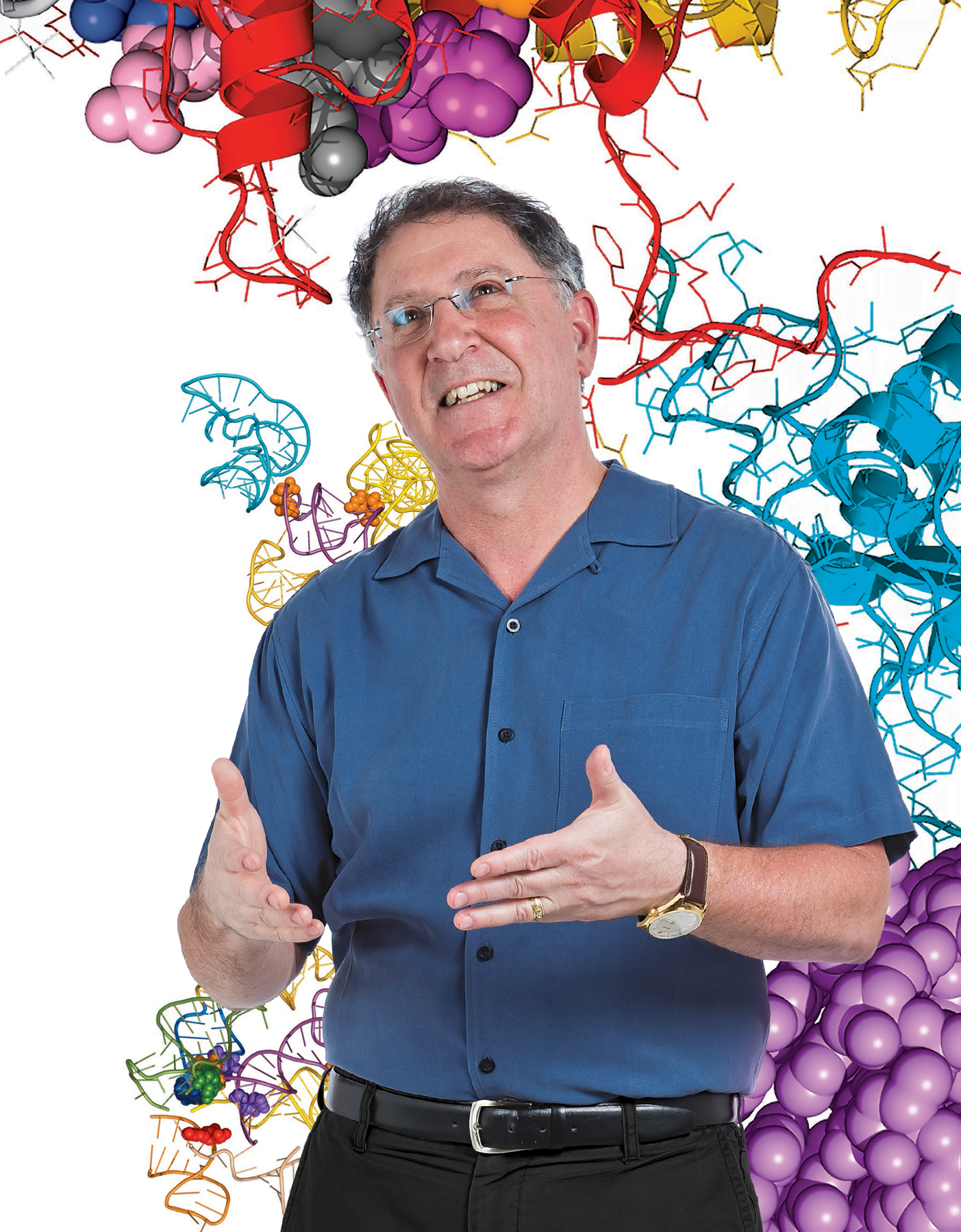
Jonathan Dinman, professor and chair of the UMD Department of Cell Biology and Molecular Genetics, has explored disease at a variety of scales throughout the course of his career.
“Some scientists start on one thing and work on that thing for their entire career. Others go where the science takes them. That’s what I’ve done,” Dinman said. “I started with parasitology. Then I became interested in yeast, then virology, which brought me to the structural biology of gene expression and human disease. That’s where I am now. I go where the interesting questions are.”
Like Dinman, diseases adapt. Genes mutate. New strains of viruses and bacteria emerge. Antibiotics and other tried-and-tested treatments lose their effectiveness. For many life scientists, staying ahead of the curve means engaging in an arms race against deadly diseases.
“Diseases are always one step ahead of us. Take the flu, for example. The virus is constantly evolving, and although we have the technology to make effective vaccines, we have to stay on top of which new strains of the virus are circulating each year. This requires a concerted global effort,” said Margaret Scull, an assistant professor of cell biology and molecular genetics. “Furthermore, as the world gets more populated and it gets easier to bounce around the globe, we’ll continue encountering new pathogens we may never have seen otherwise.”
From multispecies to the submolecular scale, UMD researchers are fighting human diseases on multiple fronts.
A Tale of Several Species
Malaria is a notorious global killer, claiming nearly half a million lives every year according to the World Health Organization (WHO). Among researchers, the disease is also notorious for its complexity: any successful anti-malaria strategy must account for the biology of three organisms—humans, mosquitoes and the parasite Plasmodium.
“Studying the malaria pathogen by itself is like listening to one hand clapping,” said Raymond St. Leger, a Distinguished University Professor in the UMD Department of Entomology who fully embraces the interdisciplinary nature of malaria research. “You need to understand all sides to make progress.”
St. Leger’s research adds a fourth layer of complexity: pathogenic fungi that specifically target insects. When these fungi come into contact with an insect’s body, the spores germinate and penetrate the insect’s exoskeleton, eventually killing the insect host from the inside out. Consequently, researchers have looked to these fungi as a way to control mosquitoes—and thus rates of malaria infection—in high-risk areas.
In collaboration with colleagues from Burkina Faso, a landlocked country in western sub-Saharan Africa, St. Leger’s group zeroed in on the fungus Metarhizium pingshaense—a natural killer of disease-carrying mosquito species including Anopheles gambiae and Aedes aegypti. To supercharge the killer fungus, the researchers genetically engineered it with several genes that express neurotoxins from spider and scorpion venom—both alone and in combination with other toxins. They published their results in June 2017 in the journal Scientific Reports.
Each engineered fungal strain killed wild-caught mosquitoes in Burkina Faso more quickly and efficiently than the unaltered fungus. But the most effective strain used a combination of two toxins already approved by the U.S. Environmental Protection Agency for insecticidal use: one derived from the North African desert scorpion Androctonus australis and another derived from the Australian Blue Mountains funnel-web spider Hadronyche versuta.
“Our most potent fungal strains, engineered to express multiple toxins, killed mosquitoes with a single spore and stopped mosquitoes from blood feeding,” said Brian Lovett, an entomology graduate student working in St. Leger’s lab and a co-author of the study. “This means that our fungal strains can prevent disease transmission by more than 90 percent of mosquitoes after just five days.”
When St. Leger, Lovett and their colleagues inserted the toxin genes into M. pingshaense, they included a highly specific promoter sequence, or genetic “switch.” This fail-safe ensured that the toxin genes could only be activated in the blood of insects and could not be released into the environment. To further ensure the safety of non-target insect species, the researchers tested the engineered fungal strains on local Burkina Faso bees. After two weeks, no bees had died as a result of the toxin-boosted fungus.
“The toxins we’re using are potent, but totally specific to insects. They are only expressed by the fungus when in an insect. Additionally, the fungus does nothing at all to bees and other beneficial species,” St. Leger said. “So we have several different layers of biosecurity at work.”
The need in Burkina Faso is dire: according to the WHO, malaria is omnipresent throughout the country and the parasite has grown resistant to the antimalarial chloroquine. The mosquitoes there have also grown resistant to traditional insecticides, according to local scientists.
“Many in Burkina Faso are very keen on this technology,” St. Leger said. “The WHO has identified insecticide resistance as the major threat to effective mosquito control, which is relevant not only to malaria but to a number of mosquito-borne diseases such as dengue and yellow fevers, viral encephalitis and filariasis. Unlike chemical insecticides that target only sodium channels, many spider and scorpion toxins hit the nervous system’s calcium and potassium ion channels, so insects have no pre-existing resistance.”
St. Leger and his collaborators plan to expand their on-the-ground testing regimen in Burkina Faso. One goal is to determine the best way to combine genetically engineered fungi with other interventions, including chemical insecticides, to prevent the evolution of resistance. Eventually, the team hopes to deploy their engineered fungal spores over a wide area to test their impact on malaria transmission.
Racing Against the Clock

While malaria requires humans, mosquitoes and parasites to exert its deadly effects, a rare genetic disease called progeria requires only a single person. But it impacts every cell in that person’s body.
Progeria mimics the normal aging process at an accelerated rate. Symptoms typically appear within the first year of life, and individuals with the disease develop thin, wrinkled skin; fragile bones and joints; full-body hair loss; and organ failure, among other complications. Most individuals with the disease do not survive past their teen years.
“Progeria affects nearly every system of the body at the cellular level,” said Kan Cao, an associate professor of cell biology and molecular genetics at UMD who studies the disease. “It is especially hard on cardiovascular cells, and the skeleton never gets a chance to develop properly.”
A defect in a single gene causes progeria. The gene produces a protein called lamin A, which sits just inside the nucleus of every cell in the body, under the nuclear membrane. Healthy cells snip off a small piece of each new lamin A molecule—a small edit that is necessary for lamin A to work properly. Cells with progeria, however, skip this important editing step. The defective lamin A interferes with the nuclear membrane, causing the nucleus to form bulges and deformations that make normal functioning impossible.
One of Cao’s earliest efforts involved the drug rapamycin, an immune suppressor commonly used to prevent the rejection of transplanted organs. She and her collaborators found the drug capable of reversing some of the cellular-level symptoms of progeria, while clearing out some of the defective lamin A proteins. But the cells still retained many of the defects associated with progeria.
Next, the researchers identified another compound that showed even more promise for treating progeria symptoms: methylene blue. In a 2015 study in the journal Aging Cell, Cao and her colleagues suggested that this common, inexpensive and safe chemical once used to treat urinary tract infections and other conditions could also be used to treat progeria. Small doses of methylene blue reversed many of the most damaging cellular-level symptoms of progeria, including misshapen nuclei and mitochondria, the small organelles that produce energy for the cell.
“We tried very hard to examine the effect of methylene blue on all known progeria symptoms,” Cao said. “It seems that it rescues every affected structure within the cell. When we looked at cells treated with methylene blue, it was hard to tell that they were progeria cells at all. It’s like magic.”
Cao and her team are moving quickly to complete the next crucial step: testing in animal models.
“So far, we have done all of our work in stem cell lines. It is critical to see whether the effect extends to whole animals,” Cao explained. “Further down the line, other groups might begin human clinical trials.”
Cao wants to extend her discoveries about progeria to the processes of normal aging. Here again, methylene blue shows promise. In a 2017 study in the journal Scientific Reports, Cao and her colleagues found that methylene blue could slow or reverse several well-known signs of normal aging when tested in cultured human skin cells and three-dimensional simulated skin tissue.
“Our work suggests that methylene blue could be a powerful antioxidant for use in skin care products,” Cao said. “The effects we are seeing are not temporary. Methylene blue appears to make fundamental, long-term changes to skin cells.”
Cao’s team also developed a three-dimensional skin model system, which her team used for further testing of methylene blue. The results suggested that methylene blue can increase skin’s thickness and ability to stay hydrated, while causing little to no irritation, even at high concentrations. Encouraged by these results, Cao hopes to develop safe and effective ways for consumers to benefit from the properties of methylene blue.
“We have already begun formulating cosmetics that contain methylene blue. Now we are looking to create marketable products,” Cao said. “We are also very excited to develop the three-dimensional skin model system. Perhaps down the road we can customize the system with bioprinting, such that we might be able to use a patient’s own cells to provide a tailor-made testing platform specific to their needs.”
Sentries at the Gates of the Human Body

Relatively speaking, whole-body diseases like progeria are an exception to the rule, because most diseases affect a single organ or a few organ systems in the body. The flu, for example, exploits vulnerabilities in one organ system—the respiratory system—to find its way past human defenses.
UMD Cell Biology and Molecular Genetics Assistant Professor Margaret Scull, who joined the department in 2016, studies the defense mechanisms that the human respiratory system uses to exclude most invaders, including the flu virus.
The respiratory system is an impressive defender of human health. Simple at first glance, but complex and multifaceted on closer inspection, many of the respiratory system’s defenses lie in the epithelial cells. These delicate cells line the inner surface of the lungs and form an interface between the external environment and the interior of the human body.
“The innate defenses present in our airway are fascinating to me,” Scull said. “The airway’s epithelial cells are an important barrier against—but also a target for—viral infection. When a virus invades your lung, epithelial cells can sense the intruder and sound the alarm, sending out biochemical signals to prepare neighboring cells for battle.”
Airway epithelial cells also have physical mechanisms that help protect us. Cells with hairlike projections called cilia line our respiratory tract, and the orchestrated movement of these cilia creates an “escalator” that can clear unwanted visitors out of the airway.
“I’m very interested in how these biochemical and physical defenses play out under different conditions,” Scull added.
It is difficult to study the dynamics of airway infection in living humans—the protocols would be far too invasive and the number of variables would complicate the analysis. Studies on cultured cells grown in the lab are also of limited value.
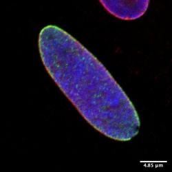
As a result, Scull uses a three-dimensional model of human airway epithelium, which allows her to investigate the activity of the ciliated cells that can physically force out invaders. The model can also highlight how different cell types in the epithelium respond to viruses. To construct the model, Scull begins with human lung cells grown in a plain cell culture dish. Next, she carefully arranges and treats the cells to encourage them to differentiate, or develop into the different types of cells that form the epithelium in human airway tissues.
“Years ago, this model was the top-of-the-line system, but there were limitations,” Scull said. “For example, if you culture cells on plastic for too long, they lose their ability to differentiate.”
Scull says that some newer systems use stem cells, the extraordinarily versatile cells that can differentiate to form any cell type in the body. Other techniques can reprogram adult lung cells to temporarily behave like stem cells and gain the ability to self-renew.
“Now we are getting to the point where we can knock out genes and pursue other more involved approaches,” Scull said. “These new models have expanded our toolbox and enabled us to finally tackle some basic questions about how airway cells fight against the flu.”
And not a moment too soon for Scull, who is eager to move on to more involved investigations. Her next target is the interaction between viruses and mucin molecules—relatively enormous proteins that form the protective mucus that coats our airway.
“It might sound funny to say it, but mucins are very dear to my heart. There is so much more to them than you might think,” Scull said. “They are massive glycoproteins with lots of sticky ends. Some are released into the airway, but others are tethered to airway cells. They clearly have a role in physically clearing pathogens, but they also seem to impact inflammation. There is so much to uncover in how mucin molecules modulate the pathogen response to keep out diseases like the flu.”
Translating the Source Code
For many hereditary diseases, researchers dive deeper than organ systems, directly targeting the defective genes in a person’s DNA. But for Jonathan Dinman, professor and chair of the UMD Department of Cell Biology and Molecular Genetics, a person’s RNA holds much more promise as a research target.
If DNA is the “hard drive” that contains all of our genetic information, then RNA—DNA’s close cousin—is more like the files and programs that do the meaningful work. The simplified model goes something like this: DNA contains genes, which a cell copies into RNA molecules. Then the cell reads the RNA to create proteins.
“The cool thing about RNA is that it’s very structurally dynamic. DNA is double stranded, which limits the possibilities,” Dinman said. “RNA is single stranded and much more flexible. It can loop back in on itself. It can combine with other RNA molecules. It can interact with proteins. The combinatorial possibilities are much broader.”
Much of Dinman’s research centers on a clever mechanism called programmed ribosomal frameshifting (PRF), which allows RNA to pack more information into a single small molecule. By prompting the cell to read the RNA molecule two different ways, one RNA molecule carries the code for two proteins. In effect, it is much like the data compression algorithms that computers use to package large files.
Many viruses that cause human disease—including HIV, Zika and chikungunya—use RNA as their genetic material instead of DNA. These viruses also use PRF to carry more information. Dinman studies how PRF works in RNA viruses and whether PRF can be exploited to defeat certain diseases.
The simple answer appears to be yes. Dinman recently looked at PRF in the context of Venezuelan equine encephalitis virus (VEEV), which is an unforgiving killer of horses, donkeys and zebras. Mortality rates reach as high as 80 percent among animals infected with the virus, and the virus can also infect humans. The U.S. and Soviet Union both weaponized VEEV during the Cold War, prompting the Centers for Disease Control and Prevention and the National Institutes of Health to classify VEEV as a category B pathogen.
Dinman and his colleagues created a mutant version of VEEV with a disrupted PRF mechanism. Tests in cultured cells did not reveal a large difference in the rate of virus production. But when the researchers tested the mutant virus in mice, they saw a dramatic increase in the rate at which infected mice survived the disease. Dinman and his colleagues published the results in the Journal of Virology in November 2016.
“With some simple mutations, we compromised VEEV’s ability to be virulent,” said the study’s lead author Joe Kendra, a biological sciences graduate student at UMD. “This result shows that PRF might be a therapeutic target for other viruses. If we can confirm that the mutant virus confers immunity—opening the door to a vaccine—that will be very exciting.”
The possibility of using PRF to defeat a number of dangerous viruses encourages Dinman. He also strongly suspects that PRF is widespread among all life forms, noting that viral models provide the best opportunity to dig deeper.
“When you want to study basic mechanisms like PRF it’s hard to do so in animals—there is so much going on. It’s much easier in viruses because the signal-to-noise ratio is better. If we look at the history of discovery, these mechanisms are always found in viruses,” Dinman said. “Science is about finding needles in haystacks. Smart scientists look in smaller haystacks.”
A Spoonful of Sugar (Molecules)
Lai-Xi Wang, a professor of chemistry and biochemistry at UMD, searches through even smaller haystacks. Much of his research program focuses on the role that molecules known as glycoproteins play in the body’s defenses against disease. Antibodies, the foot soldiers of the immune system that identify invading viruses and bacteria, are complex and highly specific glycoproteins. An antibody’s sugar groups play a large role in determining its function.
Due to subtle, yet important, differences in the structure of sugar groups, two otherwise identical antibodies might not be equally good at recruiting immune cells to kill an invading pathogen. To address this issue, Wang and his collaborators developed a method to modify an antibody’s sugar group structure and create antibodies with consistent sugar groups.
In March 2017, Wang and his colleagues published a study in the Proceedings of the National Academy of Sciences that went a step further, determining which specific sugar combinations enhance or suppress an antibody’s ability to signal the immune system to attack an invader. Even in antibodies currently used for disease therapy, a given dose might contain a wide variety of antibody variants, known as “glycoforms,” distinguished by their sugar groups.
“Our first major step forward was to develop a method to produce homogeneous glycoforms,” Wang said. “With this, we can now look at how individual different sugars affect the properties of antibodies. Until this study, we didn’t have an efficient way to know how individual sugars in various glycoforms affect suppression or activation of the immune response.”
The ability to engineer consistent glycoforms excites Wang because he says it will open a lot of doors to new therapies. Biochemists could generate highly specific therapeutic antibodies to target cancer cells and encourage the body’s immune system to kill off tumors. Erythropoietin—the hormone that prompts the body to generate new red blood cells—could be engineered to treat anemia more efficiently. And Wang’s lab is already looking at one particularly high-profile application: the prevention and treatment of HIV.
“HIV has developed very strong defensive mechanisms. One of them is glycosylation, the chemical manipulation of sugar groups,” Wang explained. “The virus is smart—it uses glycosylation to mimic the human body’s own sugars, thus fooling the immune system into mistaking the virus for a cell from the body. But there are some unusual structures in these sugars. They are not perfect replicates of the sugars found on our own cells.”
In recent years, researchers have discovered a number of neutralizing antibodies that can target these unusual sugar structures. Wang’s lab is in the process of identifying structures like these on the sugars that HIV uses to mimic human cells. This is a necessary first step, Wang said, and could enable the design of custom antibodies capable of neutralizing the virus consistently and efficiently. This, in turn, may one day provide a template for vaccine design.
“Much HIV vaccine research has attempted to target the proteins on the surface of HIV. But proteins can evolve very quickly—this is another defense mechanism. All attempts to target proteins alone have failed so far,” Wang said. “One exciting new direction in HIV vaccine and immunotherapy is to target the sugar-protein junction, called the glycopeptide epitope. Going without these sugars is not an option for the virus—it will not survive. So in theory, the virus won’t be able to escape a line of attack that targets these sugars.”
Looking Ahead
UMD researchers are optimistic regarding the future of disease research. But the push and pull of conflicting forces in the arms race against disease leaves some room for uncertainty.
“We have come so far in our ability to contain infection and create therapies, but new diseases and new epidemics emerge frequently,” Wang said. But, he notes that the fight against disease—especially major epidemics like HIV—has many dimensions that can’t be captured by one discipline alone. “HIV is not just a biomedical problem. It’s a socio-economic problem. It has far-reaching effects on social stability and economics. So it’s hard to address the issue using science alone.”
St. Leger can relate to the socio-economic dimensions of disease research. Working to fight malaria on the ground in Burkina Faso, he realized that social and cultural considerations are important pieces of the puzzle as well.
“Some people don’t want to use the cloth netting that we use to apply the spores inside their homes. Others are concerned they might be affected by the fungus,” St. Leger explained. “We get a lot of feedback and want to make sure all parties are involved. We never downplay people’s concerns. We listen and think about the best way to answer with evidence.”
While climate change and increases in population and global mobility are widely expected to enhance the spread of disease, the research community stands well poised to counter with ingenious solutions at all scales, from multispecies to submolecular.
Dinman, for his part, is bullish on the future of disease research. He thinks that the rapid pace of technological innovation coupled with the advanced state of human knowledge has put the human race in a good position.
“I think we are at a tipping point when it comes to human disease. We’ve now amassed the tools and understanding to do meaningful bioengineering,” Dinman said. “We’ve also become adept at making new tools very quickly. Tackling a disease might one day be as simple as walking into a hardware store to get what you need for a renovation project.”
Written by Matthew Wright
See also:
- Disease-fighting Alumni: A sidebar highlighting alumni whose research aims to improve the lives pf people living with autoimmune diseases, HIV and breast cancer.
- A Tale of Two Bacteriology Alumnae: A sidebar about two women whose long and successful careers began with the degrees they earned at the University of Maryland in the 1930s and 1940s.
- College Boasts Four NIH Life Science Training Grants: A sidebar about new grants that will support students studying virology and host-pathogen interactions.
This article was published in the Fall 2017 issue of Odyssey magazine. To read other stories from that issue, please visit go.umd.edu/odyssey.